
1) Describe insect integument with well labeled diagram and mention its functions.
Structure of insect integument
It consists of three parts i.e.
1. cuticle
2. epidermis or hypodermis and
3. basement membrane.
1. Cuticle: It is a complex, non-cellular layer which forms the outermost investment of the body and its appendages. The cuticle has two sub divisions viz.,
1. epicuticle (Non- chitinous) and
2. procuticle with chitin-protein complex.
1. Epicuticle: It is a very thin layer as compared to procuticle. Its thickness varies from 1/100 to 4 micron. This layer consists of three layers. A. protein epicuticle consisting of protein and polyphenol B. lipid epicuticle (wax layer) C.
tectocuticle (Cement layer). The wax layer imparts water conserving property.
2. Procuticle: It is much thicker than epicuticle. It is composed of chitin protein and other substances. Chitin is the chief constituent forming 20-50% dry weight of the cuticle. his highly resistant to alkalies. It is divisible into outer exocuticle and inner endocuticle The exocuticle is hard and dark in colour due to tanning. The endocuticle is not involved in tanning.
II Epidermis: It is one cell thick and underlies the cuticular part. Some of the cells are modified to form sense organs. It is the cellular layer of the integument that underlies and secretes the cuticle. It also produces moulting fluid, absorb digested products of the old cuticle and repairs the wound. Certain hypodermal cells produce hair like organ called setae
III Basement membrane: The inner ends of the hypodermal cells are bound by a more or less distinct membrane called basement membrane. It is 0,5 micron thick.
2) Enlist different types of mouthpart observed in insects with example. Explain the mouthparts of Cockroach & Honey bee along with well- labeled diagram.
1. Biting and chewing type: e.g. Cockroach & grasshopper.
i. Labrum
ii. Labrum-epipharynx
iii. Mandibles
iv. Maxillae
v. Hypopharynx
vi. Labium /lower lip
2. Piercing and sucking / hemipterous / bug type e.g. Plant bugs.
3. Piercing and sucking / dipterous / mosquito type : e.g. Female mosquito
4. Chewing and lapping type : e.g. honey bee.
5. Rasping and sucking : e.g. Thrips
6. Mandibulosuctorial type : e.g. grub of antlion
7. Sponging type : e.g. House fly
8. Siphoning type : e.g. Moths and butterflies
i. Labrum : (Upper lip) It is flap like, bilobed and attached to the clypeus by an articular membrane. It is movable. It covers the mouth cavity from above. It helps to pull the food into the mouth. It holds the food in position so that mandibles can act on it. It forms the roof of the pre oral food cavity.
ii. Labrum-epipharynx: Inner surface of the labrum is referred to as epipharynx. It is frequently membranous and continuous with the dorsal wall of pharnyx. It is an organ of taste.
iii. Mandibles: There is a pair of mandibles. They are the first pair of jaws. They are also called as primary jaws or true jaws. Mandibles articulate with the cranium at two points. They are heavily sclerotised. They are toothed on their inner border. There are two types of teeth. Distal are sharply pointed and are called incisor or cutting teeth and proximal teeth are called molar or grinding teeth. They act transversely to bite and grind the food into small fragments.
iv. Maxillae: They are paired and more complicated than mandibles. They are called secondary jaws or accessory jaws. At proximal end the first sclerite cardo joins the maxilla to head. The second sclerite is called stipes which articulates with cardo. Stipes carries a lateral sclerite called palpifer which bears a five segmented antenna like maxillary palp. On the distal end of the stipes, there are two lobes. The outer lobe is called galea and inner lobe is lacinia which is toothed. Maxille direct the food into the mouth. They hold the food in place when the mandibles are in action. They act as auxillary jaws and assist in mastication of food. Sense organs connected with the perception of touch, smell and taste are abundantly found in palpi.
v. Hypopharynx : It is a tongue like organ. It is located centrally in the preoral cavity. Salivary gland duct opens through it.
vi. Labium /lower lip: It is a composite structure formed by the fusion of two primitive segmented appendages. It bounds the mouth cavity from below or behind. It forms the base of the preoral cavity. It consists of three median sclerites viz., submentum (large basalsclerite), mentum (middle sclerite) and prementum (apical sclerite). On the lateral side of the prementum there are two small lateral sclerites called palpiger bearing three segmented labial palpi. Distally prementum bears two pairs of lobes. The other pair of lobes is called paraglossae and inner pair of lobes, glossae. Both pairs when fused are called ligula.
3) Define metamorphosis. State its significance and explain types of metamorphosis.
Definition of metamorphosis: The change in the body form during post embryonic development in insects is called metamorphosis.
Types of metamorphosis with examples
1. Ametabolous: Insects undergo no metamorphosis. Development takes place through three different life-stages i.e. egg, juvenile and adult. Immatures are called Juveniles. Feeding habit and habitat of immatures and adults are same. e.g. Silverfish.
2. Hemimetabolous: It is also called incomplete or gradual metamorphosis. Life-cycle is completed in three stages viz., egg, nymph and adult. Immature forms are called nymphs. Feeding habits and habitats of immature and mature forms are same. Wings develop externally. e.g. Grasshopper, Red cotton bug
3. Holometabolous: It is also called complete or complex metamorphosis. The life-cycle is completed in four stages viz., egg, larva, pupa and adult. Feeding habits, habitats and structure of immature and mature stages are completely different. Wings develop e.g. butterfly, beetles, housefly.
4. Anamorphosis: In insects like Protura, first instar larva has only 8 abdominal segments with terminal telson. The remaining three segments are added in subsequent moults. Telson remains at terminal end.
5. Hypermetamorphosis: In insects like blister beetle the larva passes through totally instars, hence known as hypermetamorphosis.
6. Epimorphosis: In which segments and legs are not added at molts e.g. Myriopods
4) Describe the Female & Male reproductive system of cockroach.
Female reproductive system of cockroach
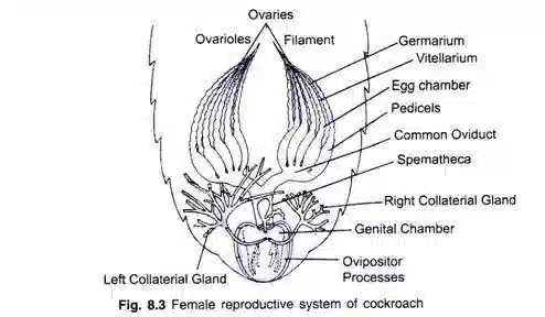
The female reproductive system of a cockroach is a relatively simple yet efficient structure that allows for the production, fertilization, and laying of eggs. As with other insects, the reproductive system in female cockroaches is composed of several key organs and structures.
1. Ovaries: The ovaries are a pair of long, coiled tubes located in the abdominal segments of the female cockroach. These structures are responsible for producing and storing the ova (eggs). The ovaries contain numerous egg chambers, or follicles, where the developing eggs are housed.
2. Oviducts: The oviducts are two narrow tubes that extend from the ovaries to the genital chamber. Once an egg is fully matured within an ovary, it is released into one of the oviducts to be transported towards the genital chamber.
3. Genital Chamber (Vagina): The genital chamber, also known as the vagina, is the terminal portion of the female reproductive tract. It receives the mature eggs from the oviducts during the process of egg-laying. Additionally, the genital chamber serves as the site of fertilization, where the sperm from a male cockroach will meet the eggs.
4. Spermatheca: The spermatheca is a specialized sac-like structure located within the genital chamber. Its main function is to store the sperm received during mating. When the female mates with a male cockroach, the sperm is transferred to the spermatheca, where it can remain viable for an extended period. This enables the female to fertilize her eggs over time without the need for repeated mating.
5. Accessory Glands: Cockroaches have accessory glands associated with their reproductive system. These glands produce secretions that are mixed with the eggs and sperm to form the egg case, or ootheca. The ootheca is a protective structure that encases the eggs and provides them with a safe environment during development. The secretion from the accessory glands hardens and forms a tough shell-like covering around the eggs.
Male reproductive system of cockroach

The male reproductive system of a cockroach is also relatively simple but efficient in its function. It consists of several organs and structures that are involved in the production and transfer of sperm to the female.
1. Testes: The testes are a pair of small, elongated structures located in the abdominal segments of the male cockroach. They are responsible for producing sperm. The sperm is then stored within the testes until it is ready to be transferred to the female during mating.
2. Vas Deferens: The vas deferens is a tube that connects each testis to the ejaculatory duct. It serves as a passageway through which sperm travels from the testes to the ejaculatory duct, where it is mixed with seminal fluid to form the spermatozoa.
3. Ejaculatory Duct: The ejaculatory duct is a short tube that receives sperm from the vas deferens. It is also the point at which seminal fluid is added to the sperm, creating the spermatozoa. The seminal fluid is produced by accessory glands associated with the male reproductive system.
4. Accessory Glands: Cockroaches have accessory glands that produce seminal fluid. This fluid contains nutrients and other substances that nourish and protect the sperm. When the seminal fluid is added to the sperm in the ejaculatory duct, it forms the spermatozoa, which will be transferred to the female during mating.
5. Aedeagus: The aedeagus is a specialized structure that acts as a copulatory organ in male cockroaches. It is used to transfer the sperm to the female's genital chamber during mating. The aedeagus is inserted into the genital chamber of the female, allowing the sperm to be released and stored in the spermatheca.

